
A. 특 징 (Features)
ㆍ병리학적인 피부 모형, 8배 확대
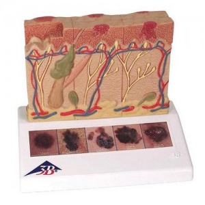
ㆍ건강한 피부와 대비되는 5가지 악성 흑색종 표현
- 건강한 피부
- 표피에 한정된 피부 표면의 암 세포
- 표피에 암 세포가 퍼져 있으며, 일부가 유두층에 침습
- 유두층에 암세포가 퍼짐
- 망상층에 암세포가 침습
- 위성 세포가 정맥과 접촉
B. 규 격 (Specifications)
14 x 10 x 11.5 cm; 0.2 kg
This 3B Scientific® Skin Pathology model shows healthy skin and 5 different stages of malignant melanoma on the front and back, enlarged 8 times:
- healthy
- malignant cells are found at the surface, within the epidermis
- malignant cells fill the epidermis, a few invade the papillary layer
- malignant cells fill the papillary layer
- malignant cells invade the reticular layer
- malignant cells have reached the subcutaneous fatty tissue, satellite cells approach a vein
In the top view of the skin cancer model, the individual stages of externally visible skin changes are shown, allowing for an assessment according to the “ABCDE” criteria. The sides of the skin cancer model show the various levels of invasion into the skin layers according to Clark (I-V) and the tumor thickness according to Breslow (in mm). 5 original color illustrations on the base of the skin cancer model show various types of malignant melanomas. The skin cancer model comes mounted on a base.
The skin cancer model is a great tool for illustrating this skin pathology.
* 무단수집및 복재를 금합니다 .
상품정보고시
| 제품명 |
피부암 모형, J15 |
| 판매가격 |
가격문의 |
| 브랜드 |
3B Scientific |
| 원산지 |
EU |
결제후 2~5일 이내에 상품을 받아 보실 수 있습니다.
국내 최대의 물류사 롯데택배를 통하여 신속하고 안전하게 배송됩니다.
30만원 이상 구입시 "택배발송" 제품에 한해서 무료배송됩니다.
(제주도를 포함한 도서,산간지역은 항공료 또는 도선료가 추가됩니다.)
택배발송 外 용달,퀵,화물비용은 구매자 부담입니다.
(글로브박스,데시게이터 등 장비류는 택배불가합니다.)
결제방법은 신용카드, 국민/BC(ISP), 무통장입금, 적립금이 있습니다.
정상적이지 못한 결제로 인한 주문으로 판단될 때는 임의로 배송이 보류되거나,주문이 취소될 수 있습니다.